Frequently Asked Questions
shortly rinsing the parenchymatous organs (spleen, liver, kidneys, etc.) in PBS or saline solution to remove blood and avoid artifacts such as hematin pigment.
fixation by immersion in 10% buffered Formalin (commercially available) for at least 24 hours at room temperature.
the volume ratio for good fixation is 10 parts formalin per 1 part tissue.
if the samples are thicker than 0.5/1 cm trimming before fixation is recommended as formalin penetrates and hence fixes the tissues with a speed of 1 mm/hour. Consequently autolytic or self-digestion processes will continue in the center of the specimen, till the fixative will be penetrating. For this reason there are references recommending fixation at 4°C temperature. Low temperature will slow down fixation but will also diminish or even blocking enzymatic activity, hence autolysis.
Some labs recommend the use of paraformaldehyde. If that’s what you have available, mind that it is not buffered and after 24 hours the pH of the solution will change with detrimental effects on epitopes and possible tissue artifact.
However the best procedure which guarantees prompt fixation and excellent quality of the histologic specimens is achieved by perfusion of the animal.
This procedure is especially indicated for investigation of the central nervous system. Additional information and protocols can be provided upon request to our email address.
In general yes it is possible even with good results. It must be considered though that not all antibodies are suitable for FFPE material and that prolonged fixation impairs partially or totally the outcome as Formalin causes denaturation of epitopes.
Poor fixation on the other hand has even a worse effect as it allows tissue and cell self-digestion with complete degradation of the epitopes.
Based on comparison of Zinc fixative vs formalin, performed at the HCF at EPFL, zinc fixative tends to shrink cells, altering often quite significantly the cyto- and histomorphology of the tissue/organ. It is however preferred by some researchers and laboratory technicians for the good outcome of immunohistochemistry when using antibodies for immune cells markers as epitopes are preserved as in frozen sections.
It is the collection of all organs and tissues of the body for histology preparation and microscopic analysis. Organs such as liver, spleen, heart and brain can be weighed and bones like the tibia measured in length and compared to the reference control. It is usually performed on genetically engineered mice to assess the morphologic impact of the gene mutation.
Because subclinical infections can influence and often impact quite severely the morphologic and/or clinical phenotype of the animal or of the experimental model.
Sure! Our team of veterinary pathologists can support you.
Our joint team of comparative and human pathologists can tell you if and to which extent your model recapitulates the disease in humans.
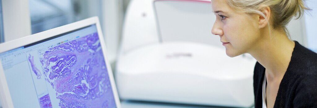
Like any other scientific data produced in research, histopathology scorings should be “objective” and reproducible.
Yes, we can design and produce customized ngTMA in which a wide array of tissue from the different species of interest can be screened simultaneously in a cost and time efficient manner. We can also set up and validate the immunohistochemistry protocol, perform the staining and analyze the results for you